 Microfluidics chambers can be used for biomedical research and development. Unlike conventional flat microplates, these do not have solid walls, so optical clarity is excellent. Besides, these chambers have the advantage of preventing "edge effects" that prevent cells from being visible. This phenomenon occurs due to the fluid dynamics that prevents cells from being located close to pinning lines. This results in improved monoclonality and the ability to pick growing clones earlier than in conventional plates. Visit xonamicrofluidics.com for application of microfluidics chambers.
The microfluidic system maintains the liquid flow in the specimen chamber. It consists of ultrathin windows and silicon microchips. The chamber is placed in a specimen holder, which seals the sample in the electron microscope. It also incorporates tubing to and from the sample, which is connected to a syringe pump outside the microscope. We characterised the liquid flow by performing fluorescence microscopy on spheres made from gold nanoparticles. We then imaged the gold nanoparticles using a 200 kV STEM. Upon establishing a stable cell culture, a microfluidics chamber can be used for research. There are several applications for this technology, including the detection of toxins, the analysis of DNA sequences, and the creation of inkjet printing devices. You can read a detailed review about its uses here. So, if you're interested in microfluidics chambers and how they can benefit your research, you can check out these applications. The microfluidic chamber is a versatile tool for neuroscience research. It can be used to grow a variety of cells, such as neurons and tumors. It can also be used for studying the interaction between cells and other tissues. As the chamber is small, it is perfect for a single experiment. Moreover, it can be used to investigate the behavior of a large number of different tissues, and can be a great tool for drug discovery. Dry etching is a technique that is used to transfer patterns to a substrate, but it is expensive and requires a lot of time. A biotin-containing microfluidics chamber is the ideal solution for the research. These biofabrics chambers allow researchers to monitor a range of biological processes and identify the mechanisms that control them. These devices also allow for the manipulation of complex molecules, such as DNA. Kindly go to website for adequate info on microfluidic chamber. The microfluidics chamber is an ideal instrument for analyzing the chemistry of complex fluids and microbial cells. These labs use the chambers to analyze the behavior of cells under various conditions. Xona Microfluidics, LLC provided the chambers used in this research. The American Heart Association and the National Institute of Mental Health funded the project. The team would like to thank the Xona Microfluidics company for providing the chambers. Besides microfluidics chambers, microfluidics circuits can also be used to create pores. The size of the pores can be as small as 100 nm, allowing for more efficient and accurate research. The cells were rolled on the surface of the E-selectin-coated microfluidic chamber and fixed using a fixation solution containing 3% paraformaldehyde and 0.2% glutaraldehyde. For more information, check out this related post: https://en.wikipedia.org/wiki/Microfluidics .
0 Comments
 Microfluidics chambers have become increasingly popular due to their versatility. Despite their size, a single-cell array can be constructed within a minute. In addition, they can be used for a range of other applications, from imaging cancer cells to analyzing the function of the brain. They are an ideal tool for a wide variety of research purposes. In this article, we discuss some of their advantages.
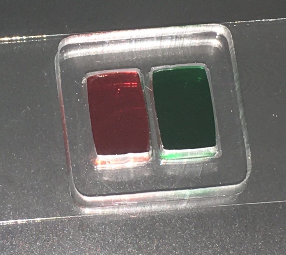
A microfluidics chamber is unique because its fluid walls allow for excellent optical clarity. Conventional flat microplates tend to have walls that trap cells against the edges, leading to "edge effects," which make it difficult to see cells clearly. However, this is not the case with a microfluidics chamber. In these vessels, incoming cell-containing medium forces preexisting cell-free medium toward the sides. The resulting condition allows for much higher visual clarity, which helps users to determine whether a cell is monoclonal. It is also possible to select a clone earlier than in a conventional plate. Visit https://xonamicrofluidics.com/product/rd150/ to find out the advantages to using microfluidics. A microfluidic chamber is ideal for studies requiring high-throughput, and high-resolution image analysis. In addition, microfluidics can be used for detection of toxins, DNA sequencing, and creating inkjet printing devices. These systems are a useful tool for a wide variety of scientific fields. The aqueous phase, which is normally pinned to the Petri dish, is reshaped by a Teflon stylus, resulting in an array of individual chambers. The liquid walls of FC40 isolate each chamber from its neighbors, preventing the formation of microfluidic structures. A microfluidic chamber allows for precise measurement of individual cells in a single cell. The researchers were experimenting with a technique to measure individual cells in worms. The microfluidic chamber allowed them to observe muscle cell inactivity during certain periods. Inactivity is not the same as sleep! The researchers were able to measure each individual cell with a syringe and see if it was active or inactive. Read this article for more details on a microfluidic chamber . A microfluidic chamber is also suitable for cell culture. It allows scientists to control the amount of medium, which is essential for performing accurate measurements. This technology also allows researchers to manipulate the cells' movements. As a result, the biofluidics chamber can be used for toxicity testing and to grow cells. The use of a microfluidic chamber increases the speed and flexibility of laboratory experiments. These devices can also be used for the analysis of many different biological samples. A microfluidic chamber is useful for a variety of research purposes. It provides a high-resolution environment for cell culture. The microfluidic chamber is made of molded elastomeric polymer that fits on a glass coverslip. The axonal and somal compartments are separated by a physical barrier with embedded microgrooves. This makes it possible to study the function of different cell types in a complex manner.Check out this related post to get more enlightened on the topic: https://en.wikipedia.org/wiki/Microfluidic_cell_culture .  There are several distributors that offer xona software microfluidic devices, but it's not always easy to figure out which one is right for your needs. Most of these devices are custom-made for specific applications, and they need certain geometries, inlet/outlet sizes, and channel depths. The best place to buy microfluid devices is a distributor that doesn't sell pre-made chips, because that way, you'll have complete intellectual property protection and the best quality device. The microfluidic chip is a great example of one. These devices are small and compact enough to fit into a pocket, making them extremely convenient to carry around. You can even store a sample in one. And if you're using it in your lab, you can keep it in a safe place so you can keep your samples safe. Many of these microfluidic chips can be used in research settings as well, so they're a great investment for your lab. Another popular microfluidic device is the Lab-on-a-Chip. These devices work by placing very small amounts of fluid onto a microchip. They are great for performing tests and diagnosing diseases, such as Alzheimer's disease, ALS, and Parkinson's. These devices can also be used for other purposes. These devices are made to be portable and cost very little. But there are some downsides to them, too. If you are unsure, consider buying a kit from a manufacturer that has a warranty. The first type of microfluidic device is the Lab-on-a-Chip. It is a device with a spiralling channel, and it is called a lab-on-a-chip. It is a device that has branches on its outside. The flow enters at the center of the device and exits at the outer end. These devices are designed for particle separation based on inertia. They are useful for separating particles of different shapes, sizes, and weights. The number of branches will depend on the application. The market for microfluidic devices is growing worldwide. China and India are expected to have the fastest growth. The region's economy is booming and the market is expected to increase rapidly over the next few years. There are several applications for microfluidics, including detecting toxins, testing DNA sequences, and inkjet printing. For more information, visit the dedicated review page of the industry. These are just a few of the reasons to buy microfluid devices. Check out this page to get more information on microfluidic devices. The market for microfluidic devices is growing globally. The demand for these devices is driven by the availability of a wide range of products at reasonable prices. The majority of these products are made of glass or plastic. The price of these devices is high, and the cost is rising. Most of these devices are manufactured with a high quality and precision, and can even be customized to your specifications. However, it is important to choose the best supplier for your particular application.If you want to know more about this topic, then click here: https://en.wikipedia.org/wiki/Microfluidics_in_chemical_biology . |
AuthorWrite something about yourself. No need to be fancy, just an overview. ArchivesCategories |
 RSS Feed
